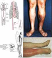
DEEP VEIN THROMBOSIS Deep vein thrombosis, also called deep venous thrombosis or DVT, is a blood clot (thrombus) in a deep vein. It usually affects the leg veins, such as the femoral vein or the popliteal vein.
Deep-vein thrombosis occurs in about 1 in 1000 people per year. About 1-5% will die from the complications of a DVT like pulmonary embolism (lung blood clots). Dr Virchow noticed an association between venous thrombosis in the legs and pulmonary embolism. Heparin, a type of anticoagulant medication that keeps the blood thin and thus prevents blood clots, was introduced to clinical practice in 1937. In the last 25 years, considerable progress has been made in the understanding of, diagnosis and treatment of deep vein thrombosis DVT. There may be no early symptoms of a DVT. Some people may have a DVT clot without even realizing it as the DVT clots are small and not significant. However when there are symptoms, the most common symptoms are of pain and swelling of the leg or calf. Read on to find out more about DVT symptoms. What causes a DVT ? There are many factors that increase the likelihood of a clot forming. The main factors are immobilization, recent trauma or surgery and cancer. Read on to find out more about DVT causes. Deep vein thrombosis treatment is mainly centered around preventing new clots from forming with blood thinners and allowing time for the body to break down the clot by itself. In rare situations like very large DVT clots, medications are used to break down the DVT but this carries with it and increased risk of bleeding. Read on to find out more about DVT treatments. Diagnosis Venography uses a radiographic material injected into a vein on the top of the foot. The material mixes with blood and flows toward the heart. An X-ray of the leg and pelvis will then show the calf and thigh veins and reveals any blockages. Although venography is very accurate and can detect blockages in both the thigh and the calf, it is also costly and cannot be repeated often. In addition, the injected material may actually contribute to the creation of thrombi. Duplex ultrasonography can also be very accurate in identifying clogged veins. Projected sound waves bounce off structures in the leg and create images that reveal abnormalities. The addition of color Doppler imaging improves accuracy. This test is noninvasive and painless, requires no radiation, can be repeated regularly and can reveal other causes for symptoms. It also costs substantially less than venography. However, it is technically demanding and requires a skilled, experienced operator to obtain the most accurate results. Ultrasonography is less sensitive in detecting thrombi in the calf and it has limited ability to directly image the deep veins of the pelvis. Magnetic resonance imaging is particularly effective in diagnosing DVT in the pelvis, and as effective as venography in diagnosing DVT in the thigh. This technique is being increasingly used because it is noninvasive and allows simultaneous visualization of both legs. However, an MRI is expensive, not always readily available, and cannot be used if the patient has certain implants, such as a pacemaker. In addition, the patient can experience claustrophobia. |
HOPES PINNED ON HIM
People from all over the world queue to see acupuncturist Leong Hong Tole who has made a name for himself in the world of traditional complementary medicine...Read Source: The Star Newspaper (Malaysia) |
| :: CLICK HERE FOR MORE VIDEO :: |
QUESTION |


